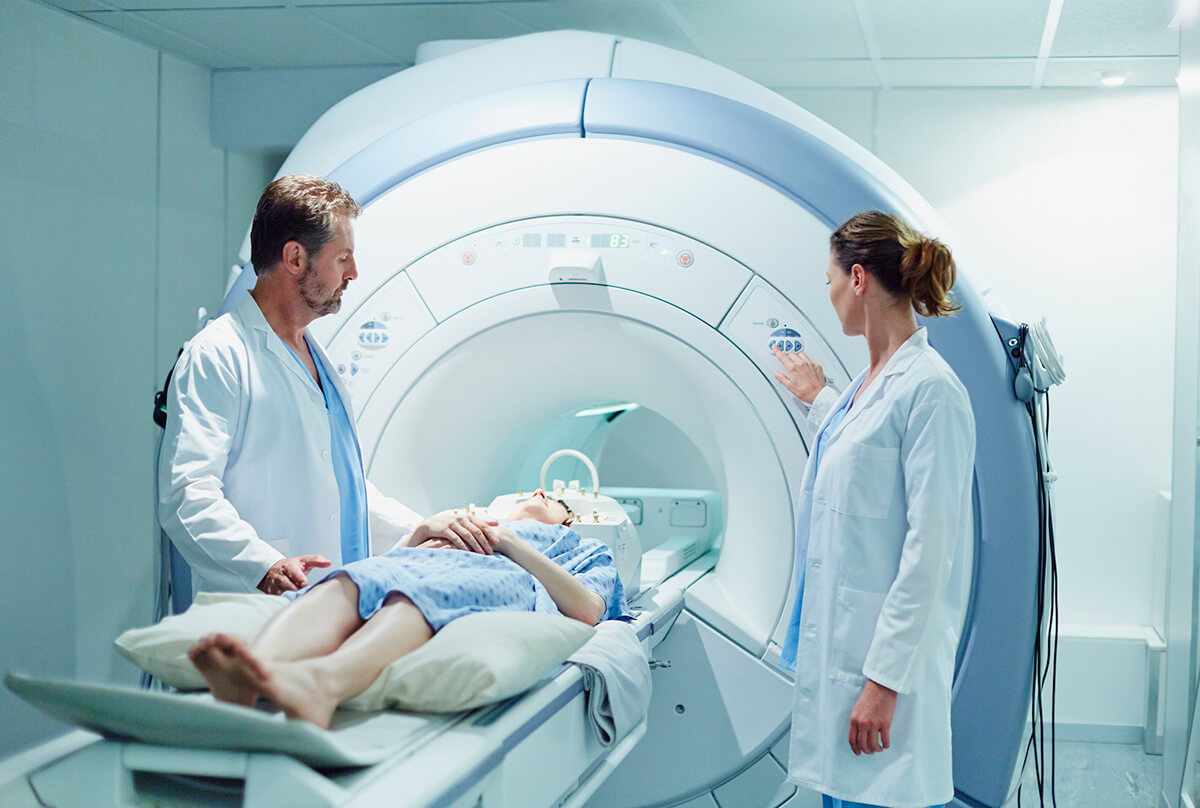
Magnetic resonance imaging is a modern non- X-ray method for imaging various organs, parts and structures in the body. It is one of the best ways to diagnose musculoskeletal, central nervous and endocrine diseases. This method is based on the principle of radio waves forming a magnetic field, which are directed to the desired part of the body in order to visualize its parts. The method is harmless for adults and particularly suitable for children because there is no harmful irradiation.
This type of imaging provides the most detailed information about the body, in the presence of injuries and lacerations, problems with bones, cartilage, tendons. The most common application of MRI is the diagnosis of brain tumors, strokes and cardiac pathologies. Thanks to it, it can provide very early detection of a number of conditions so that their treatment can be far more effective. The excellent quality of magnetic resonance images can provide the best possible information if surgical intervention is subsequently required.
Magnetic resonance imaging provides detailed information on the location, size and reactivity of pathological tissue changes in malignant diseases.
Why is an MRI done?
MRI is a method of high diagnostic value, especially suitable for patients who need to have several consecutive examinations in a short time and avoid X-rays. Since the introduction of MRI into practice, many diseases do not require biopsy because the test detects and diagnoses a wide range of pathological conditions at an early stage. It can provide very early detection of a range of conditions so that treatment can be far more effective.
In medical practice, MRI is also used to differentiate tumor from normal tissue, to diagnose organs of the chest and abdomen, including:
- heart
- liver
- bile ducts
- Kidney
- spleen
- Gut
- pancreas
- adrenal glands
- pelvic organs
- including reproductive organs
- blood vessels, etc.
In sports, MRI is the primary method for diagnosing the musculoskeletal system and soft tissues, i.e. muscles, joints, bones, tendons, etc.
What is an MRI scan itself?
A standard examination lasts between 15 and 45 minutes, depending on the problem and the organs for which multiple images need to be collected for diagnostic purposes. During the examination itself, the patient is in a supine position and should not move during this time. A device called a "coil" is placed next to the area to be examined. Nowadays, there are different types of MRI machines - open and closed, and scanners now offer a fairly wide space for the patient. So patients suffering from anxiety or claustrophobia will feel more relaxed. Also, during the scan itself, the patient can be accompanied by a loved one so that they feel as comfortable as possible.When the scan itself starts, knocking noises and machine buzzing are heard at different frequencies and intensities. Protecting the psyche and anxiety in some cases the patient is even played music. Certain cases in an MRI scan, necessitates the use of a chemical substance to increase the contrast of the image. It is a liquid that is injected into a vein (most often in the inner side of the elbow) and is non-iodine-containing.
When proceeding with the examination itself, it is a good idea for the patient to have previously removed any metal accessories and jewellery (piercings, earrings, glasses, watches, rings, etc.) from any part of the body. Women are advised to be make-up free because of cosmetic substances and products that would interfere with the study. In case of:
- implanted metal splints, plates, pins, artificial limbs, presence of metal particles in the body (shrapnel, bullets, shavings)
- as well as the presence of any electronic devices in the body (pacemakers, artificial heart valves)
the specialists performing the MRI examination must be notified in advance. Clothing and underwear should be of natural fabrics, as artificial fabrics degrade the quality of the images and may need to be removed.
When it is necessary to use a contrast agent is fine:
- The patient should not have eaten for at least 3 hours before the examination;
- if the patient has allergies to foods and medicines, it is desirable to consult an allergist;
- after the examination, it is advisable for the patient to take more fluids - at least 2 liters during the first twenty-four hours;
- alcohol should not be consumed after the examination;
There is no perceptible direct impact on the patient's senses during an MRI scan, apart from the sound of the machine. It varies in strength and frequency depending on the site being scanned. The examination itself is easily tolerated by patients as the magnetic field and radio waves are not felt and do not cause unpleasant sensations or discomfort.
The MRI scan is safe, poses no health risks to the patient and has no side effects.
For more information, we at Medical Karaj are at your service.
Call us on the following numbers "Medical Karaj": 0879 977 401 or 0879 977 402.


